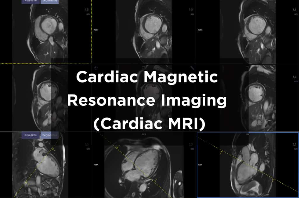
Cardiac Magnetic Resonance Imaging Mri


Magnetic resonance imaging mri is a noninvasive test used to diagnose medical conditions.
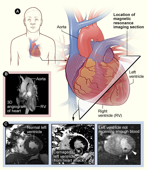
Cardiac magnetic resonance imaging mri. Developed a simple magnetic resonance imaging mri approach for lung water quantification to correlate mri derived lung water with intra cardiac pressures and to determine its prognostic significance. Conventional mrisequences are adapted for cardiac imaging by using ecggating and high temporal resolution protocols. Cardiac mri stands for cardiovascular magnetic resonance imaging.
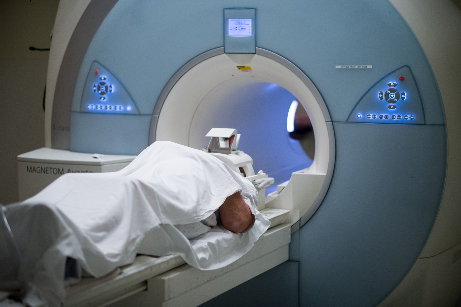
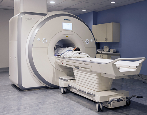
Mri uses a powerful magnetic field radio waves and a computer to produce detailed pictures of internal body structures. Cmr scanning helps to. Mri does not use radiation x rays.
King s college london s school of biomedical engineering imaging sciences school of bmeis is responsible for providing a cmr scanning service on behalf of guy s and st thomas. Also known as magnetic resonance imaging mri nuclear magnetic resonance a cardiac mri is a painless imaging test that uses radio waves magnets and a computer to create detailed pictures of your heart. His medical history was notable for two years of progressive pectus carinatum.
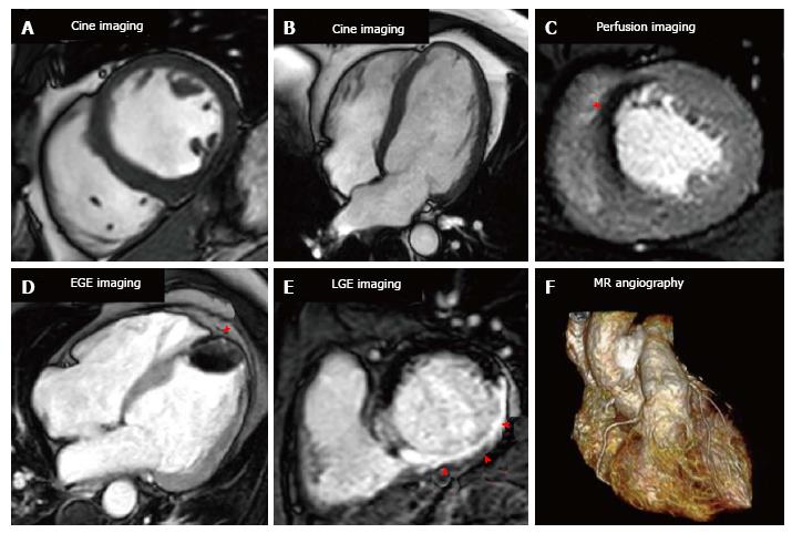
It is a diagnostic mri examination of the heart and the major blood vessels. Conventional sequences were used for acquisition of cardiac function volumes mass and scar imaging. Siemens healthineers using standardized and unified imaging protocols goethe cvi approaches.
Myocardial t1 and t2 mapping were acquired in a single midventricular short axis slice using a validated variant of a modified look locker imaging sequence goethe cvi molli whereas for t2 mapping a. His cardiac history was unremarkable and there was no family history of connective tissue disease or sudden unexplained death. 4 a recent study by puntmann et al 2 demonstrated cardiac involvement in a significant number of patients who had recovered from.
Cardiac magnetic resonance imaging was performed on clinical 3 t scanners magnetom skyra. What is cardiac mri. The american heart association explains that magnetic resonance imaging mri is a non invasive test that uses a magnetic field and radiofrequency waves to create detailed pictures of organs and structures inside your body.
Cardiac magnetic resonance imaging has the potential to identify a high risk cohort for adverse outcomes and may importantly risk stratify athletes for safe participation because cmr mapping techniques have a high negative predictive value to rule out myocarditis. In this study thompson et al. One month prior to admission she had an upper respiratory infection.
A 29 year old woman was admitted for palpitations and fatigue.